Study: Lung biopsy cryoprobe increases diagnostic yield over standard forceps
In a new study published in JAMA, the diagnostic yield of transbronchial lung biopsy was significantly higher when using a cryoprobe versus forceps in a group of patients with pulmonary nodules or masses, recent lung transplant, and diffuse parenchymal lung disease.

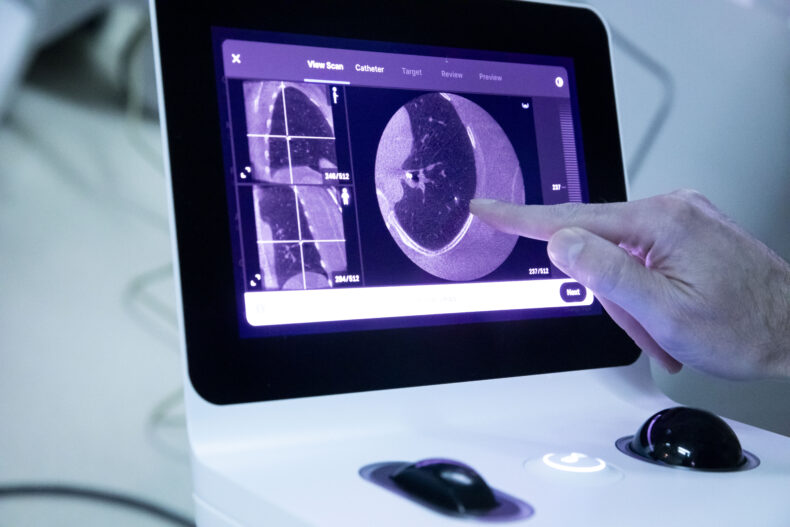
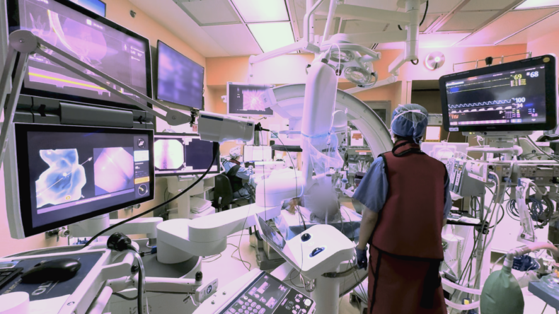
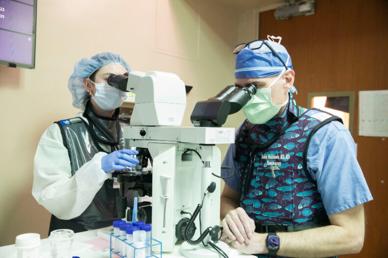
A transbronchial lung biopsy is a minimally invasive procedure in which a bronchoscope — a thin, lighted tube — is guided through the nose or mouth into the lungs. Tools are then passed through the scope to collect tissue for laboratory analysis to diagnose lung conditions. A cryoprobe is a medical instrument that uses localized freezing to extract tissue. Forceps are used to pinch off tissue for removal, which can be faster but also crushes a portion of the tissue sample.
The FROSTBITE-2 randomized trial showed diagnostic yield during transbronchial biopsy was nearly 10 percentage points higher when performed using a 1.1-millimeter cryoprobe rather than with 2.0-millimeter forceps (88.6% vs 78.8%). The difference was particularly great among patients with pulmonary nodules or masses (83.2% vs 70.1%). In a secondary safety analysis, there were four pneumothoraces (collapsed lungs) requiring chest tube placement in the forceps group (1.6%) compared to none in the cryoprobe group. No patients experienced significant bleeding or respiratory failure events.
“A structurally intact, sufficiently large tissue sample from a targeted area in the lung increases the likelihood of an accurate diagnosis, which is what we strive for every time we perform a transbronchial lung biopsy,” said interventional pulmonologist Fabien Maldonado, MD, MSc, Professor of Medicine and Thoracic Surgery, and Director of Interventional Pulmonology at the Vanderbilt Lung Institute.
“We’re continually investigating ways we can improve these procedures, as accurate diagnoses up-front save time, which may help get patients the treatment they need faster. Evaluating the tools we use, particularly as innovations in this area occur, is an important avenue of investigation.
“Individuals who have known or suspected lung issues deserve to have the best possible diagnostic procedures, so they and their clinical teams have clear evidence of what is occurring in their lungs so informed treatment decisions can be made.”
Previous studies using a 1.9-millimeter cryoprobe have yielded larger lung tissue specimens at higher quality without crushing the sample, but there were also more bleeding and pneumothorax events. The FROSTBITE-2 trial used the 1.1-millimeter cryoprobe probe which, unlike the larger probes, is small enough to remove the biopsy specimen through the working channel without having to remove the scope, which increases safety.
Certain lots of the cryoprobe went under a Food and Drug Administration Class I recall in March due to reports of rupturing or bursting during activation; none of these events were reported in this trial.
The study was conducted under the auspices of the Interventional Pulmonary Outcomes Group, an international collaborative of clinical experts dedicated to improving patient care in interventional pulmonology through multicenter clinical trials and research. Maldonado, who holds the Pierre Massion Directorship in Lung Cancer Research at Vanderbilt Health, is vice chair of this group.
The trial was completed at nine U.S. medical centers including Vanderbilt Health that perform at least 100 transbronchial biopsies annually and have affiliated institutional centers for lung cancer, lung transplant and interstitial lung disease. Patients enrolled were 18 or older and scheduled to undergo transbronchial biopsy for lung nodules or masses, lung transplant, or diffuse parenchymal lung disease. Five hundred individuals were randomly assigned to either the 1.1-millimeter cryoprobe or the 2.0-millimeter forceps for the biopsy.
“These promising results bring us one step closer to making these vital diagnostic procedures even more safe, accurate and effective,” said Vanderbilt Health interventional pulmonologist Robert Lentz, MD, Associate Professor of Medicine and Thoracic Surgery. “Our team is currently conducting FROSTBITE-3, a randomized controlled trial comparing the 1.1-millimeter cryoprobe with instruments for lymph node biopsies, to determine whether this novel tool may help with molecular testing in patients diagnosed with lung cancer.
The FROSTBITE-2 study is an investigator-initiated trial. It was funded by Erbe, an international business that develops, manufactures and markets surgical systems. The funder had no role in trial design, data collection, data analysis, manuscript preparation, or the decision to publish.
The post Study: Lung biopsy cryoprobe increases diagnostic yield over standard forceps appeared first on Vanderbilt Health News.