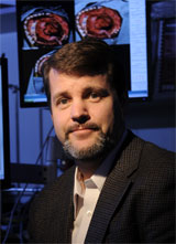
Michael I. Miga, M.S., Ph.D.

  • Harvie Branscomb Professor of Biomedical Engineering
  • Professor of Neurological Surgery
  • Professor of Otolaryngology
  • Professor of Radiology & Radiological Sciences

Phone

615-343-8336

Email

michael.i.miga@vanderbilt.edu
Vanderbilt University Medical Center
Stevenson Center
VU Station B, #351631
Nashville, TN 37235

Michael I. Miga, M.S., Ph.D.

  • Harvie Branscomb Professor of Biomedical Engineering
  • Professor of Neurological Surgery
  • Professor of Otolaryngology
  • Professor of Radiology & Radiological Sciences

615-343-8336

michael.i.miga@vanderbilt.edu

Vanderbilt University Medical Center
Stevenson Center
VU Station B, #351631
Nashville, TN 37235

Profile

Dr. Miga is an expert in computational modeling, medical image analysis, inverse problems, elastographic image reconstruction, and image-guided interventions for biomedical engineering applications. He has been in the field since 1994 and has been in professional status since 2000. He is currently director of the Biomedical Modeling Laboratory at Vanderbilt University and has multiple NIH grants. He is also a co-founder of the Vanderbilt initiative in Surgery and Engineering.

The focus of the Dr. Miga's laboratory is on new paradigms in detection, diagnosis, characterization, and treatment of disease through the integration of computational models into research and clinical practice. With the continued improvements in high performance computing, the ability to translate computational modeling from predictive roles to ones that are more integrated within diagnostic and therapeutic applications is becoming a rapid reality. With respect to therapeutic applications, efforts in image-guided surgery applications in brain, liver, kidney, and breast tumor resections are being investigated. His work centers around the use of imaging data for the execution of surgical procedures and in image contrast mechanisms that use mechanical properties for the purposes of guiding clinical applications, i.e. interventional diagnostics. Recent collaborative work using elastographic imaging for the characterization of low grade gliomas, breast tumors, and neoadjuvant therapeutic responses has been another area of interest. In addition to that work, tumor growth models are also being developed to assist in macroscopic characterization of disease as well as morphological changes to surrounding structure for also evaluating neoadjuvant chemotherapy responses. The common thread that ties the work together is that, throughout each research project, the integration of mathematical models, tissue mechanics, instrumentation, and analysis is present with a central focus at translating the information to directing therapy/intervention or characterizing tissue changes for diagnostic value.


Education

  • Ph.D., Dartmouth College, Hanover, New Hampshire (1998)
  • M.S., University of Rhode Island, Kingston, Rhode Island (1994)
  • B.S., University of Rhode Island, Kingston, Rhode Island (1992)
Research Emphasis

Research Description

Publications

Have any questions? Contact Us 1-877-936-8422 for more information